For dentists
CBCT, panoramic and scans for the referring dentist — in one click
Keep CBCT, panoramic, intraoral X-rays and scans in one place, and the referring colleague can view them right away in the browser — no CD burning, no courier.
Register your practice Viewing doctors: free Download the brochure (PDF)
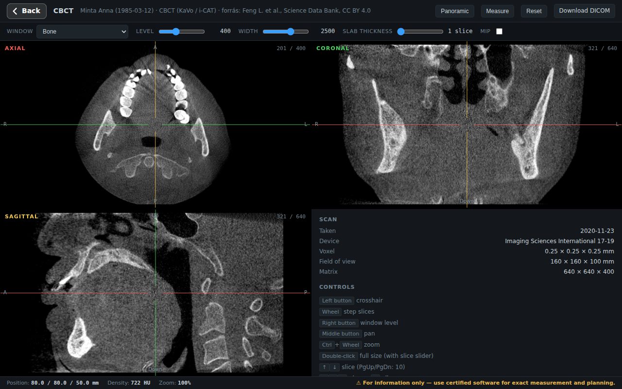
3D CBCT viewer in the browser
The CBCT opens in three planes, without installation, on computers and phones. Double-click to enlarge one plane, scroll slices with the slider or the mouse wheel.
- Dental windows: bone, teeth / enamel, airway / sinus
- Slab thickness and MIP — thin structures stay visible
- Distance measurement in mm (informational)
- The original DICOM downloads in one click

Panoramic and cross-section from the CBCT
Mark the dental arch with a few clicks on the axial image — Sinus3D reconstructs a panoramic from it. A draggable red line on the panoramic shows where you see the cross-section perpendicular to the arch.
- Panoramic in “focal layer”, average or MIP mode, adjustable slab
- Cross-section from buccal to lingual, with a 10 mm scale
- Measurement on the panoramic and the cross-section
- Step millimetre by millimetre along the arch

Panoramic and intraoral X-rays
2D images open in their original tone, with zoom and pan. A single, restrained “Enhance” slider helps with details — the original is one click away.
- Panoramic, periapical, bitewing, cephalometric, photo
- DICOM, JPG, PNG, TIFF
- No photo editing that could mislead — the image stays the original

Intraoral scans in 3D
The scanner export (STL, PLY, OBJ or ZIP) can be rotated in 3D: upper and lower arch separated, in the same position as in the mouth, or in occlusion.
- Occlusion also by the right or left bite registration (registration shown in blue)
- The scanner photos next to the model
- Rotate and zoom with mouse or finger
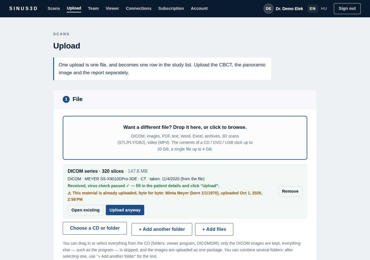
Upload: the CBCT export or the patient’s CD
Choose the file, the folder or the CD: the upload starts immediately in the background while you check the patient details. DICOM images are identified automatically; the viewer software and accompanying files are not uploaded.
- Single-file CBCT, slice series and ZIP — tested with scans from real devices
- Patient name and date of birth taken automatically from the DICOM header
- Virus scanning on our own server, without third-party services
- Duplicate detection: you are notified if the same scan is already in the system
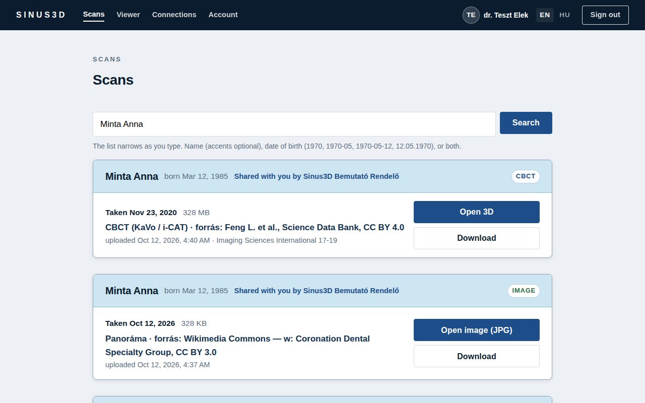
The referring dentist sees it right away
The practice and the referring colleague approve each other once. After that you just choose who the scan was made for — they are notified, and the scan appears in their list.
- Only approved colleagues see what you send them
- Sharing can be revoked at any time
- Access log: who opened what, and when
- Currently free for referring dentists

On the phone too
At a consultation, away from the practice: CBCT and X-rays open on the phone too, zoom with two fingers, double-tap to switch to one plane.
- Can be added to the home screen like an app
- Phone notification when a scan is sent
How does it work?
Upload
The practice uploads the scan — the device export, a folder or the patient’s whole CD. The patient’s name is filled in from the file.
Share
Choose who it was made for. The referring doctor is notified right away by e-mail or on their phone.
View
The referring doctor opens it in the browser, on a computer or a phone — no software, no CDs, no courier. The original DICOM can always be downloaded.
For the patient too
The patient can get an expiring link to their own scan; it opens only with their date of birth.
SSL Labs: A+ — connection encryption rated in the top category by the independent Qualys SSL Labs test (November 2026). Verify →
Frequently asked questions
Which CBCT devices are supported?
Any device’s standard DICOM export: single-file CBCT, slice series (folder or ZIP), compressed DICOM too. Tested with scans from real devices.
Does the panoramic replace a panoramic X-ray unit?
No — the panoramic and cross-sections reconstructed from CBCT are informational, for a quick overview and for discussing the case with a colleague. For diagnosis and planning, open the original DICOM in certified software.
Does the referring colleague need to install software?
No. Every viewer runs in the browser, on computers and phones; the colleague clicks the link sent to them and signs in.
How much does it cost?
Currently free for referring, viewing dentists. The practice that uploads scans pays a monthly fee from HUF 9 900 (gross) — plan and storage can be extended at any time.
Try it
Register as a practice — or as a referring doctor who views the scans sent to them: currently free of charge. After signing in, every feature can be tried on the demo practice’s sample scans.
Register your practice Working in another field? For physicians →
The viewers are informational; Sinus3D is not a medical device. For diagnosis, implant or surgical planning, open the original DICOM file in certified software. The pictures were made from public, freely licensed sample scans.