Windowing, MPR and panoramic reconstruction: what does the recipient see?
The same CT slice looks completely different in a brain window and a bone window. What windowing means, how plane reconstructions are made, and when viewing in a browser is enough.
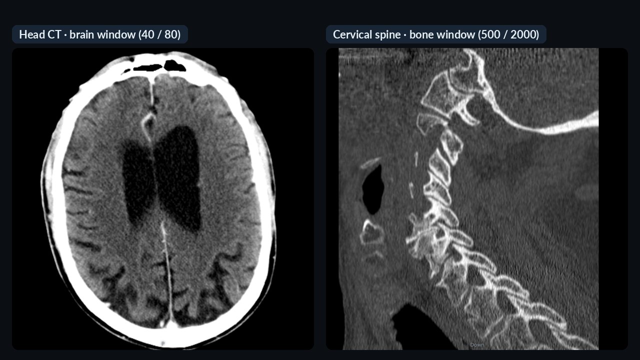
Windowing: a slice of the Hounsfield scale
CT measures the X-ray attenuation of tissue in Hounsfield units (HU): water is 0, air −1000, cortical bone around +1000. That is thousands of grey levels — far more than a monitor or the eye can separate. So a viewer always shows a window: a range of a given width around a centre (level), spread from black to white. Anything below is black, anything above is white.
| Window | Centre / width (HU) | For |
|---|---|---|
| Brain | 40 / 80 | brain parenchyma, haemorrhage |
| Soft tissue | 40 / 400 | abdomen, neck, muscle |
| Lung | −600 / 1500 | lung parenchyma, airways |
| Bone | 500 / 2000 | bone structure, fractures |
These are typical values and vary between institutions. Hospital CT scanners also send their own suggested window in the header — a good viewer uses it as the starting view and also takes the series type (e.g. a sharp bone kernel) into account.
CBCT is different: dental CBCT grey values are not calibrated HU. There the window only helps visibility; density (e.g. bone quality) cannot be measured reliably from the grey value.
MPR: any plane from the volume
CT, CBCT and 3D MR series describe a volume. Multiplanar reconstruction (MPR) computes any plane from it: the usual axial, coronal and sagittal planes, or oblique ones. Slab thickness matters: a thin slice shows more detail but more noise; a thicker (averaged or MIP) slab makes vessels or root canals easier to follow.
Curved plane: a panoramic view from CBCT
In dentistry, the image “straightened out” along a curve drawn on the dental arch is a curved planar reformation. The result is a panoramic-like image with perpendicular cross-sections. It is excellent for overview and patient information, but it has limits: its accuracy depends on the position of the curve and the slab thickness, it can distort, and it does not replace a true panoramic radiograph or measurements in certified planning software.
Diagnostic or informational viewing?
| Diagnostic | Informational | |
|---|---|---|
| Data | the original DICOM | the original DICOM or a viewing copy of it |
| Software | certified as a medical device | browser or phone viewer |
| Environment | suitable, calibrated monitor and reading room | anywhere |
| Purpose | report, measurement, planning | consultation, “look at this region”, patient information |
Under the EU Medical Device Regulation (MDR, 2017/745), software intended to support diagnostic or therapeutic decisions is a medical device (Annex VIII, Rule 11). Systems for storing, transferring and informational viewing are typically not — this is decided by the intended purpose defined by the manufacturer, and the interface should say so (“not for diagnostic use”).
In practice, the point of sharing is that the recipient gets the original data. A quick browser preview complements it — for diagnosis, download the original and open it in your own certified software.
Frequently asked questions
What is the difference between a brain window and a bone window?
They show the same CT data in different ranges: the brain window (40/80 HU) reveals subtle differences in brain tissue, the bone window (500/2000 HU) shows bone structure. Detail visible in one becomes black or white in the other.
Can bone density be measured on CBCT?
Not reliably: CBCT grey values are not calibrated Hounsfield units and vary between devices and settings.
Does a panoramic reconstruction from CBCT replace a panoramic radiograph?
Not fully: it is good for overview, but its accuracy depends on the drawn curve and slab thickness, and it can distort.
May I diagnose in a browser viewer?
A browser viewer is fine for information. For diagnosis, reporting and measurement, open the original DICOM in software certified as a medical device.